CMU Study Uncovers How Dangerous Bacteria Build and Break Down Biofilms
High-throughput, label-free method reveals more than 100 genes tied to pneumococcal biofilm development
Heidi Opdyke
- Associate Dean of Marketing and Communications, MCS
- Email opdyke@andrew.cmu.edu
- Phone 412-268-9982
Researchers at Carnegie Mellon University have developed a powerful new approach to studying how bacteria form and dissolve biofilms — complex, multicellular communities that play a central role in infectious disease. The method is already providing new insights into Streptococcus pneumoniae, also known as pneumococcus, a leading cause of death from respiratory infections worldwide.
Pneumococcus can live harmlessly in the back of the nose or throat without causing symptoms, but sometimes the bacteria spread to other parts of the body, leading to illnesses that range from mild to severe.
“We operate from the hypothesis that understanding how pneumococcus transitions from an asymptomatic colonization to a symptomatic infection will open new avenues for treatment,” said Luisa Hiller, associate professor of biological sciences.
While they can exist as single cells, bacteria typically live in complex communities called biofilms.Within these communities, bacteria display collective behaviors such as cooperation, division of labor, and competition. Because biofilms contribute to persistent and severe infections, they pose a major challenge in medicine. Understanding how biofilms form and function is critical for both basic science and the development of new therapies.
While Hiller’s group has identified some of the signaling molecules used by pneumococcus, a comprehensive system to study its biofilm development had been lacking.
The new work, published in mBio, reveals the framework to generate a comprehensive, life cycle-level perspective of individual pneumo cells as they move through various stages of the biofilm life cycle.
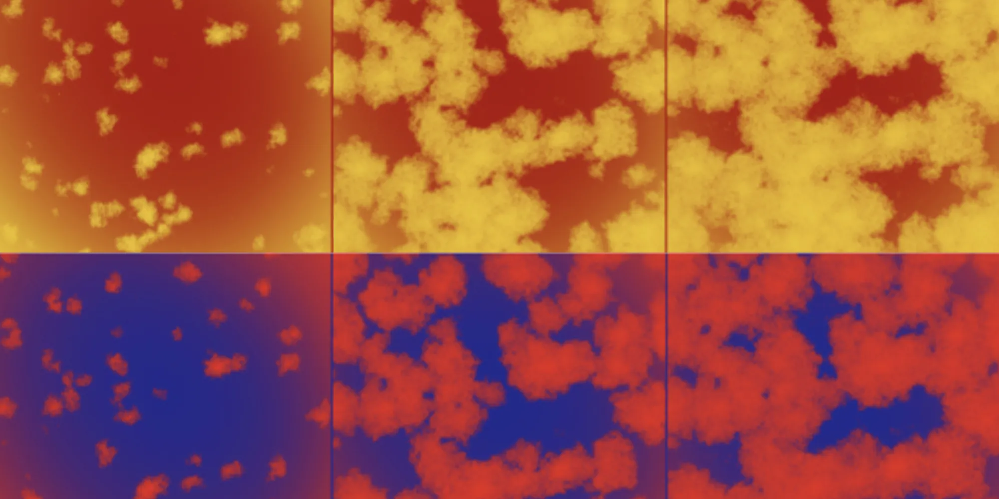
“An important advance that we present in this study is the ability to visualize individual cells growing into microcolony biofilms, which provides the granularity required to observe various stages of the biofilm life cycle,” said graduate student Pratyush Madapuji Ravi. “This, when combined with being able to process hundreds of samples in a day, enables us to screen for genes that affect various stages of the biofilm life cycle, including formation, maturation and dispersal.”
The latest research is a collaboration between the Hiller lab and the lab of Assistant Professor of Biological Sciences Drew Bridges, who studies Vibrio cholerae, the bacterium that causes cholera. Bridges’ lab previously developed a screening platform to analyze genes and behaviors across the biofilm life cycle.

“Our platform can be used to inform researchers of the genes that underlie the biofilm regulation of diverse species and could be used to identify new biofilm-disrupting drugs,” Bridges said. “Simultaneously, this paper really lays out the fundamental genetics underlying the specific pathogen S. pneumoniae.”
Graduate students Madapuji Ravi and Jojo Prentice adapted the Bridges platform to work across multiple species, with a particular focus on pneumococcal biofilms. Their innovation, called Label-Free Analysis of Biofilms (LFAB), allows researchers to observe the entire biofilm life cycle — from initial surface attachment to growth into microcolonies and eventual dispersal — without disrupting the system. LFAB requires relatively low-cost equipment, captures both spatial and temporal dynamics, and enables high-throughput screening.
Using LFAB, the researchers analyzed more than 2,000 pneumococcal genetic mutants and identified over 100 genes with potential roles in biofilm development. Notably, many of these genes had not previously been linked to biofilm formation, opening new avenues for discovery.
This life cycle-level perspective fills a major gap in the field.

The findings lay the groundwork for future research aimed at understanding how genes, signaling networks, and environmental factors interact to shape biofilm development. The researchers are now using LFAB to study how stressors — such as antibiotics — affect biofilm growth, structure, and behavior.
For Madapuji Ravi, the work also opens new directions for his thesis.
“Namely, I want to answer how S. pneumoniae has evolved resistance to antibiotics by horizontal gene transfer,” Madapuji Ravi said. “Biofilms are the sites where this species naturally exchanges DNA with its neighbors and evolves. Therefore, having now established a biofilm model, I can study horizontal gene transfer and the evolution of antibiotic resistance within biofilms, a question of high clinical importance.”
For Ravi, the work also opens new directions for his thesis.
“Namely, I want to answer how S. pneumoniae evolves resistance to antibiotics by horizontal gene transfer,” Ravid said. “Biofilms are the sites where this species naturally exchanges DNA with its neighbors and evolves. Therefore, having now established a biofilm model, I can study horizontal gene transfer and the evolution of antibiotic resistance within biofilms, a question of high clinical importance.”
Ultimately, the researchers aim to create a comprehensive map linking genes to biofilm behavior over time and space. Such insights could reveal new vulnerabilities in bacterial communities and guide the development of therapies designed to disrupt biofilms and combat persistent infections.
This research was supported by a range of federal, foundation, and institutional funding sources. Support included grants from the National Institute of Allergy and Infectious Diseases (R00AI158939) and the National Institute of General Medical Sciences (1R35GM160020), as well as a Shurl and Kay Curci Foundation grant, a Kaufman Foundation New Investigator Research Grant (KA2023-136488), and a Damon Runyon Cancer Research Foundation Dale F. Frey Award for Breakthrough Scientists (2302-17) awarded to A.A.B., along with startup funds from Carnegie Mellon University. Additional support came from NIH grant R21AI182784 and a Kaufman Foundation New Initiative Research Grant (KA2024-144002) awarded to N.L.H., a fellowship from Carnegie Mellon’s Center for Machine Learning and Health at the School of Computer Science supporting P.M.R, and the Department of Biological Sciences at Carnegie Mellon University.