Lymphocyte Telomere Length (PCS3, subsample)
Funding for measurement of lymphocyte telomere length was not included in the original PCS3 grant (NIAID R01 AI066367), but instead was obtained through a supplement awarded 3 years into the course of the study (NCCAM RC1AT005799; see Supporting Grants). Accordingly, blood samples for examination of telomere length were collected beginning with Trial 4 and continued through to the end of Trial 13. A final subsample of 152 participants provided blood samples with sufficient genetic material to assess telomere length for at least one of the 4 cell populations of interest (see below).

Sample Collection
Whole blood for telomere assay was collected from a random 1/3 of the sample on each of 3 days: during the baseline screening visit (6-8 weeks before quarantine); 3-5 days before quarantine; or on the baseline day of quarantine. 15-ml samples were collected by standard venipuncture into 3 green-top (heparinized) collection tubes. Blood was drawn from only a subsample of participants at a time because we did not have resources available to conduct timely cell separations and preservation for all participants in a single day.
Lymphocyte Sorting
Lymphocytes sorting was performed in whole blood using flow cytometry (2 ml aliquot each for antibody labeling and cell counts). Peripheral blood mononuclear cells (PBMCs) and serum were separated following a protocol of Ficoll-Paque™ PLUS (Cat# 17-1440-03, Amersham Biosciences, Pittsburgh, PA)
Lymphocyte subpopulations were separated from whole blood using the RoboSep™ automated cell separator (STEMCELL™ Technologies). CD4+ cells were isolated using the EasySep® Human Whole Blood CD4 Positive Selection Kit (18082); and CD19+ cells using the EasySep® Human Whole Blood CD19 Positive Selection Kit (18084). CD8+ cells were separated using the RosetteSep® Human CD8+ T Cell Enrichment Cocktail (15063); and CD28+/CD28- cells using the EasySep® Human PE Positive Selection Kit (18551).
Assay
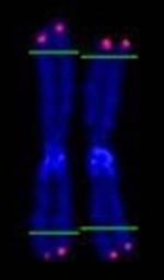
Mean telomere length was measured in peripheral blood mononuclear cells (PBMCs), CD4+ cells, CD19+ cells, and two subpopulations of CD8+ cells (CD8+CD28+ and CD8+CD28-) using a real-time quantitative polymerase chain reaction (qPCR) assay that determines the relative ratio of telomere repeat copy number to single-copy gene copy number (T/S ratio) in experimental samples as compared with a reference DNA sample following a published protocol1.
All samples were run in duplicate, with replicate values being averaged to obtain the final T/S ratio values for use in analyses. Final T/S ratio values for a given cell type were computed only for those participants with complete data for both replicates. Participants missing one or both replicate values for a given cell type were assigned a missing final T/S ratio value for that cell.
Reference
1 O’Callaghan, N.J., Dhillon, V.S., Thomas, P., & Fenech, M. (2008). A quantitative real-time PCR method for absolute telomere length. BioTechniques, 44, 807-809.