Noninvasive Neuromodulation
Transcranial focused ultrasound
Transcranial focused ultrasound (tFUS) is becoming an attractive neuromodulation tool due to its high spatial selectivity through a non-surgical and low-energy procedure (He, 2016). Low-intensity tFUS has demonstrated these merits through animal and human studies by targeting itself at cortical/deep brain regions. Despite of the rapid increase of the tFUS being used in neuroscience research, there is still unmet need in documenting the brain areas that are activated/inhibited by this mechanical energy. Our lab is developing a non-invasive EEG-based neuroimaging tool (electrophysiological source imaging, ESI) with a natural setting in order to register the area and measure the acoustic-induced neuromodulatory effects. Animal models have been employed to successfully test this tFUS-ESI concept (Yu et al. 2016). Our initial study shows that it is possible to image brain electrical activity from noninvasive scalp EEG recordings following tFUS perturbation. Our results suggest that ESI may become a useful tool to derive biomarkers (besides motor response and fMRI reading) to quantify tFUS effects and guide its use for neuromodulation. Such noninvasive biomarker, while obtained in real time, may offer important insights to optimize tFUS stimulation parameters and make tFUS a closed-loop neuromodulation modality. Human study is also underway to further test this low-intensity tFUS-ESI approach.
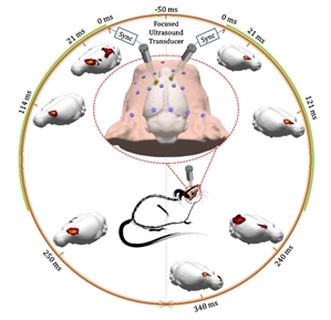
Electrophysiological source imaging of brain networks perturbed by low-intensity transcranial focused ultrasound using a wild-type rat model (from Yu et al., IEEE Trans. Biomed. Eng., 2016)
Transcranial magnetic stimulation and electroencephalography
Transcranial magnetic stimulation (TMS) is a form of noninvasive neuromodulation that can perturb neural activity or temporarily modulate excitability in focal areas of the cortex. By combining TMS with neuroimaging approaches, we are exploring several exciting avenues of research. In particular, we are focusing on the integration of electroencephalography (EEG) and TMS, to image ongoing electrophysiological dynamics and changes in activity in response to TMS. This work aims to enhance the reliability and specificity of TMS as an interventional tool, and to improve our understanding of connectivity between different network nodes in the brain. We have shown that identical TMS pulses delivered to primary motor cortex can elicit different evoked potentials and network activity as measured by EEG, depending on whether the pulse elicited a suprathreshold motor response (Petrichella et al., 2017). Additional work in the lab has looked at approaches integrating TMS interventions with brain-computer interface practice for enhanced motor rehabilitation after stroke (Johnson et al., 2017).
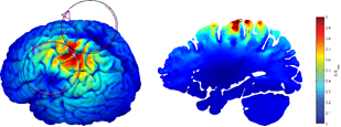
Electric field induced in the brain by TMS, as predicted by a computational model. (from Cline et al., IEEE-EMBC, 2015)