Characterizing electrode microstructures
X-ray CT scans are used every day to diagnose medical problems. How can they help diagnose transport issues in porous electrodes?
We use nanoscale CT scans of a porous electrode to give new insight into its 3D microstructures. With knowledge of an electrode's physical structure, we can form a stronger understanding of transport in the electrode.
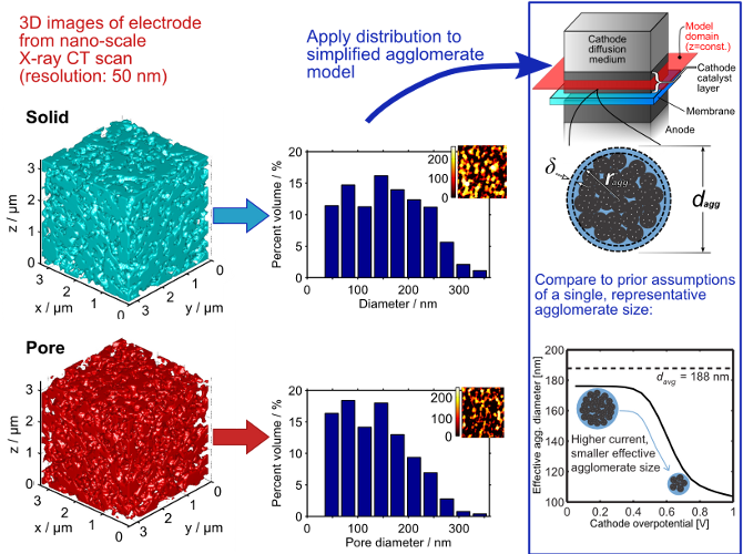
The porous electrode of a polymer electrolyte fuel cell has a 3D microstructure that is dictated by its composition and the conditions of its formation. Typically, we describe it as being made up of "agglomerates" of platinum/carbon catalyst particles bound together by ionomer (a proton-conducting polymer). Between those agglomerates are macro-pores. With greater knowledge of the 3D microstructure of the electrode, we can examine how parameters like porosity, tortuosity, agglomerate sizes, and pore sizes affect transport in the electrode. Furthermore, with a means to measure the 3D microstructure, we can learn how different electrode fabrication techniques can affect those parameters.
We use nano-scale X-ray computed tomography to examine the 3D microstructure. This technique is like a medical CT scan, but the resolution is much higher (50 nm) and the contrast is strong enough to distinguish e.g. carbon (agglomerates) from air (pores). We have validated this technique using other established techniques like electron microscopy and porosimetry.
Using the microstructural images from this technique, we have also examined a popular mathematical model of a fuel cell electrode. Typically, this model uses a single, representative agglomerate size. We learned that using instead a realistic distribution of multiple agglomerate sizes has a significant effect on the results.
For more information, check out this brief explanation of our microstructural characterization (PDF) or the following items:
- Journal paper: W. K. Epting, J. Gelb, S. Litster, “Resolving the Three-Dimensional Microstructure of Polymer Electrolyte Fuel Cell Electrodes using Nanometer-Scale X-ray Computed Tomography,” Advanced Functional Materials, 22, pp. 555-560 (2012). [PDF][Supplementary PDF][News]
- Journal paper: W.K. Epting, S. Litster, "Effects of an Agglomerate Size Distribution on the PEFC Agglomerate Model," Int. J. Hydrogen Energ., 37, pp. 8505-8511 (2012). [PDF][Supplementary PDF][News]
- Conference video: W. K. Epting, S. Litster, J. Gelb, "Nanoscale X-ray Computed Tomography of Polymer Electrolyte Fuel Cell Electrodes," Meeting of the Materials Research Society, Boston, MA, Dec. 2, 2011. [Video and news item on web]
- Conference paper: S. Litster, K.C. Hess, W.K. Epting, and J. Gelb, “Catalyst layer analysis: Nanoscale X-ray CT, spatially-resolved in-situ microscale diagnostics, and modeling,” Transactions of the Electrochemical Society, Boston, MA, Oct. 10, 2011. [Paper on web]