Epting et al: Nano-CT of PEFC Electrode Published

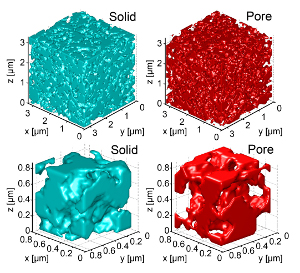 3D images of the microstructure of a PEFC electrode, reconstructed by nanoscale X-ray CT with a resolution of 50 nm.
In November 2011, Advanced Functional Materials published a paper on X-ray CT characterization of the 3D microstructure of a polymer electrolyte fuel cell (PEFC) electrode at 50 nm resolution. The work, titled "Resolving the Three-dimensional Micro-structure of Polymer Electrolyte Fuel Cell Electrodes using Nano-scale X-ray Computed Tomography," was produced by TPES lab members Billy Epting and Prof. Shawn Litster, as well as Jeff Gelb, a collaborator from Xradia, Inc.
The paper describes not only the nanoscale X-ray CT (nano-CT) measurements, which use Zernike phase contrast to achieve a 50 nm resolution in imaging these porous electrodes that are mostly carbon, but also a rigorous validation of the results. Analysis of mercury intrusion porosimetry and transmission electron microscopy measurements confirmed that the nano-CT's 50 nm resolution was sufficient for imaging the porous structure of the electrode.
Subsequently, image analysis yielded spherical size distributions from the pore and solid phases, and the volume-average solid diameters were found to be 188 nm and 222 nm in the two different samples studied. Such data will be useful to PEFC researchers for e.g. evaluating electrode preparation, or obtaining parameters for computational and analytical electrode models.
The paper is available in Advanced Functional Materials with DOI 10.1002/adfm.201101525.