| |
Research Highlights:
Precise Structures
HIV-1
Cholesterol
Peptides in Membranes
Rafts
Water Permeability Through Membranes
|
|
Questions: How can we detect membrane microheterogeneity (Rafts) without using a fluorescent probe? Is wide-angle X-ray scattering (WAXS), with a resolution of a few angstroms, sensitive to liquid-liquid phase coexistence (the hallmark of membrane Rafts)? What about Bragg D-spacings of coexisting phases? What can we learn about orientation of tie-lines in raft ternary mixtures?
 Liquid-Liquid Domains in Bilayers Detected by Wide Angle X-Ray Scattering Liquid-Liquid Domains in Bilayers Detected by Wide Angle X-Ray Scattering
Biophysical Journal Volume 95 July 2008 682–690
Thalia T. Mills1, Stephanie Tristram-Nagle2, Frederick A. Heberle3, Nelson F. Morales4, Jiang Zhao3,
Jing Wu3, Gilman E. S. Toombes1,5, John F. Nagle2, and Gerald W. Feigenson3
1Department of Physics, Cornell University, Ithaca, New York 14853; 2Department of Physics, Carnegie Mellon University, Pittsburgh, Pennsylvania 15213; 3Field of Biophysics, and 4Department of Chemistry, Cornell University, Ithaca, New York 14853; and 5Unite´s mixtes de recherche 168 Centre National de la Recherche Scientifique/Institut Curie, 75005 Paris, France |
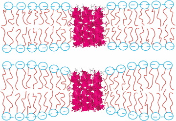
a b s t r a c t Wide angle x-ray scattering (WAXS) from oriented lipid multilayers is used to examine liquid-ordered (Lo)/liquid disordered (Ld) phase coexistence in the system 1,2-dioleoyl-sn-glycero-3-phosphocholine/ 1,2-dipalmitoyl-sn-glycero-3-phospho-choline/
cholesterol (DOPC/DPPC/Chol), which is a model for the outer leaflet of the animal cell plasma membrane. Using the method of analysis developed in the accompanying work, we find that two orientational distributions are necessary to fit the
WAXS data at lower temperatures, whereas only one distribution is needed at temperatures higher than the miscibility transition
temperature, Tmix = 25–35oC (for 1:1 DOPC/DPPC with 15%, 20%, 25%, and 30% Chol). We propose that the necessity for two
distributions is a criterion for coexistence of Lo domains with a high Sx-ray order parameter and Ld domains with a lower order parameter. This criterion is capable of detecting coexistence of small domains or rafts that the conventional x-ray criterion of two
lamellar D spacings may not. Our Tmix values tend to be slightly larger than published NMR results and microscopy results when
the fluorescence probe artifact is considered. This is consistent with the sensitivity of WAXS to very short time and length scales, which makes it more capable of detecting small, short-lived domains that are likely close to Tmix. |
1 component - poor fit. 2 components - good fit.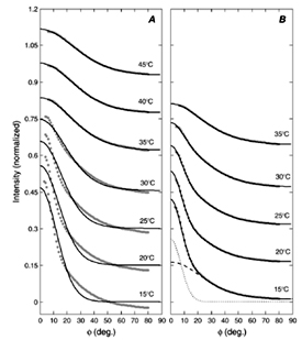 |
D-spacing alone does not always detect liquid-liquid phase coexistence.
 |
We welcome you to read the article - click here.
|
Orientation of Tie-Les in the Phase Diagram of DOPC/DPPC/ Cholesterol Model Biomembranes
Langmuir Volume 26 (2010) 17363–17368
Pradeep Uppamoochikkal, Stephanie Tristram-Nagle, and John F. Nagle
Biological Physics, Department of Physics, Carnegie Mellon University, Pittsburgh, PA
|
abstract
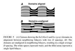
We report the direction of tie-lines of coexisting phases in a ternary diagram of DOPC/DPPC/ cholesterol lipid bilayers, which has been a system of interest in the discussion of biological rafts. For coexisting Ld and Lo phases, we find that the orientation angle R of the tie-lines increases as the cholesterol concentration increases and it also increases as temperature increases from T=15 degC toT=30 degC. Results at lower cholesterol concentrations support the existence of a different two-phase coexistence region of Ld and So phases and the existence of a three-phase region separating the two two-phase regions. Our method uses the X-ray lamellar D-spacings observed in oriented bilayers as a function of varying hydration. Although this method does not obtain the ends of the tie-lines, it gives precise values (+/-1deg) of their angles (alpha) in the ternary phase diagram. |
 |
 |
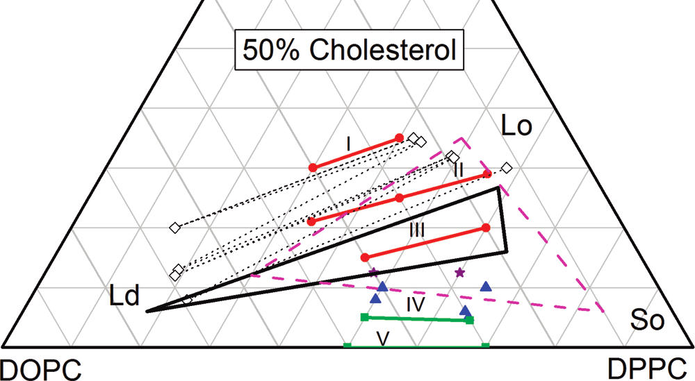
| Figure 1. NMR tie-lines of Veatch et al. are shown by dotted straight lines, and open diamonds show end points at the two-phase Ld-Lo boundary. The Ld-Lo-So three-phase region of Veatch et al. is shown as a solid black triangle, and the dashed magenta triangle is the three-phase
region from Davis et al. The orientations (not the end points) of our tie-lines are labeled I-V. The red circles and the green squares are in two-phase regions, the blue triangles are in a three-phase region, and the purple stars could be in either twophase or three-phase regions. All results are for T=15 degC except for T=18 degC for the three-phase triangle of Davis et al. |
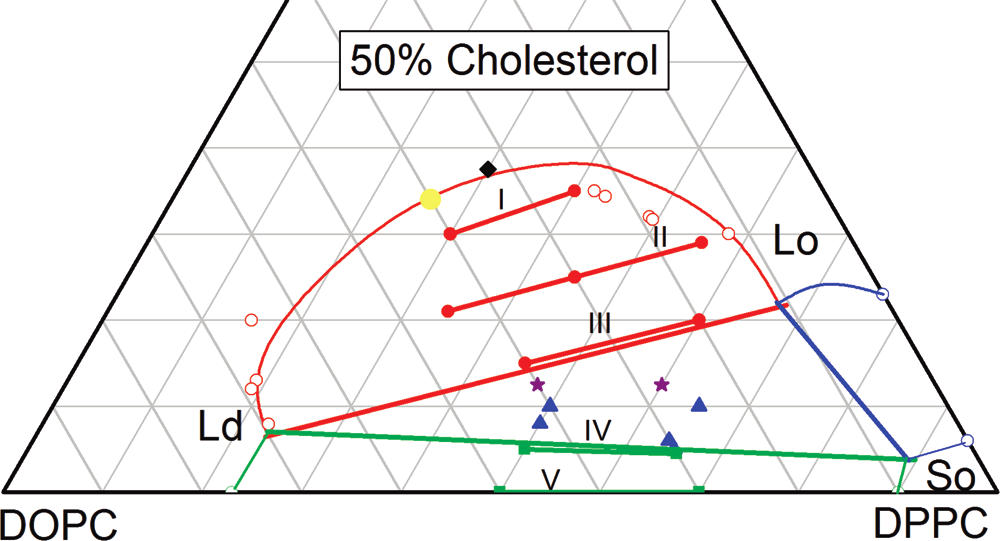
Figure 2. Suggested phase diagram at T=15 degC. The Ld-So two phase region is enclosed by three straight green lines and contains our IV and V tie-line fragments; the two open green circles at zero Chol show the coexistence compositions of Mitsui and Furuya. The Ld-Lo two-phase region is enclosed by one straight and one curved line, and it contains our I, II, and III tie-line fragments; the open red circles show the coexistence compositions of Veatch et al. The Lo-So two-phase region is enclosed by two straight lines and one curved blue line; the open blue circles at zero DOPC show the coexistence compositions of Vist and Davis. The yellow point suggests the location of an upper consulate (critical) point, and we found only one D-spacing for the composition of the black diamond. |
| We welcome you to read the article - click here. |
|
|
|
|