X-ray computed tomography
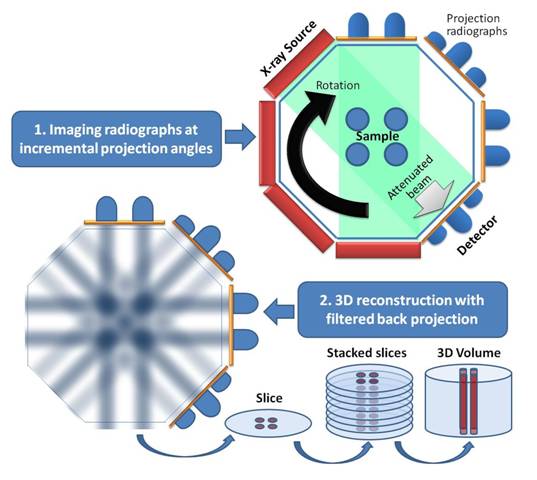
The schematic below shows the process of generating 3D, volumetric images using X-ray computed tomography (XCT). XCT is based on taking 2D ragiographs through a sample at many projections around the sample and computationally reconstructing a 3D image from the projections. The 2D radiographs are created by passing a X-ray beam from an X-ray source through the sample and imaging the resulting attenuated X-ray on the detector (a scintallator that converts X-rays to visible light coupled to a CCD camera). The X-ray attenuation is due to X-ray absorption (photoelectric absorption or Compton scattering). At the 8 keV photon energy of the XCTF's nano-CT, photoelectric absorption dominates. The amount of X-ray absorption strongly depends on the atomic number, Z, and density of the material - higher Z and higher density yielding more volumetric absorption.
Once a large enough number of projections are obtained to achieve the desired resolutions (many hundreds of projection over 180 degrees), the 3D image is recontructed using filtered back projection. The schematic below illustrates this process where the 2D radiographs (shown as 1D for simplicity) are simultaneously project back into the image space and the intersection of the back projections generates the 3D images that can be viewed as virtual slices or 3D volumes.
Additional introductory information on X-ray CT can be found on the site for the High Resolution X-ray CT Facility at UT Austin.