A team of researchers from Carnegie Mellon and the University of Pittsburgh received a five-year, $13.3 million grant from the National Institutes of Health (NIH) to establish a National Technology Center for Networks and Pathways, a new research facility that has the potential to create new technologies to unlock fundamental aspects of diseases such as cancer, dementia and stroke.
The center, headquartered at Carnegie Mellon, focuses on developing fluorescent probe and imaging technologies to investigate regulatory pathways and networks in real-time in living cells. This work generates powerful molecular biosensors for preclinical research to map the many cell-signaling networks involved in disease. Ultimately, such biosensors will be used in hospital- and office-based diagnostic medicine, which improve healthcare worldwide.
"The award recognizes our visionary science and the collaborative strength of both universities in advancing an exciting new field," said Mark Kamlet, provost and senior vice president at Carnegie Mellon. "The major grant gives us even greater capabilities to produce innovative biotechnologies that impact tomorrow's medicine."
"The further development of discrete intracellular fluorescent probes is the critical next step in exploiting the results of the Human Genome and Human Proteome projects," said physician and scientist Arthur S. Levine, senior vice chancellor and dean of the School of Medicine at the University of Pittsburgh. "The combined research resources and research faculty of the two universities promise to move the whole of medical science and medicine forward with an exceptionally exciting and new momentum."
Within any given human cell, hundreds, if not thousands, of proteins may interact in precise cascades when the cell is called upon to yield a necessary product, such as a hormone, or to respond to an environmental stress, according to center faculty. Understanding the elements and dynamics of these cascades in a given cell and in real-time is the key to diagnosing why cells don't function properly and what might be done about that dysfunction therapeutically.
"The tools created at the center give us a completely new way of looking at complex biological processes, allowing us to watch these activities in unprecedented ways," said Norka Ruiz Bravo, deputy director for Extramural Research at NIH.
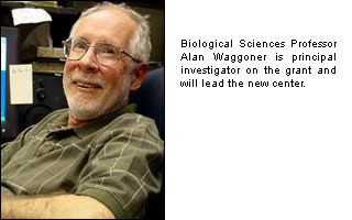
"We are poised to build on an exciting, productive foundation of tools developed at Carnegie Mellon," said Alan Waggoner, principal investigator of the grant and director of the new center. Waggoner directs Carnegie Mellon's Molecular Biosensor and Imaging Center (MBIC), which for more than 20 years has designed fluorescent probes used worldwide for research, ranging from the study of nerve activity to understanding gene activation as part of the Human Genome Project. "The grant from NIH puts us on the map and recognizes our vital collaboration with the University of Pittsburgh and other renowned academic partners to design, image and test a new generation of probes in living systems," he said.
The grant was one of three awarded as part of "Building Blocks, Biological Pathways and Networks Roadmap for Medical Research,” an NIH initiative that aims to support research that develops innovative tools that determine in real-time the amounts, locations and interactions of large numbers of individual proteins within a single cell. Such research stands to produce the next generation of research tools required to fully understand cell networks and how they function in health and disease. The NIH Roadmap is a series of far-reaching initiatives designed to transform the nation's medical research capabilities and speed the movement of research discoveries from the bench to the bedside.
"A cell's regulatory pathways are very complicated, and we need to learn so much more. But to date there are almost no tools available to sort out all of these different interactions and activities that are going on in cells," said Waggoner. "We're developing a powerful toolbox of intracellular fluorescent biosensors that cells themselves will produce. With these tools, we can study in detail how all the proteins are interacting with each other in real-time in the 3-D space of a living cell."
Effectively using these intracellular fluorescent probes and biosensors requires an array of current and new detector technologies, according to Simon Watkins, co-director of the Center for Networks and Pathways. Much of the imaging work is carried out at the University of Pittsburgh's Center for Biologic Imaging (CBI), widely recognized as one of the premier imaging centers in the country. The CBI focuses on the application of fluorescent microscopy techniques and their translation to the study of living cells and organisms.
Fluorescent probes under design at Carnegie Mellon include integrated molecular biosensors, which are being developed through a series of unique, high-throughput technologies pioneered by Carnegie Mellon scientists.
"The new center truly revolutionizes how we witness certain catalytic activities inside living cells," Waggoner stated. "Until now, no one has created fluorescent probes to visualize kinase and phosphatase reactions. But there are many hundreds of these reactions within cells, and they are critical to driving pathways involved in disorders like cancer, diabetes, stroke and infection."
The Carnegie Mellon team has world-renowned expertise in biochemistry, genetics, dye chemistry and imaging. Their current work stems from a 35-year history of developing thousands of powerful fluorescent probes that have been widely commercialized and that have made a profound impact on biomedical research.
Related Links:
"Building Blocks, Biological Pathways and Networks Roadmap for Medical Research"
Molecular Biosensor and Imaging Center