SCD Project - Pain, cerebral autoregulation, and cognition
Sickle cell disease (SCD) is a debilitating disease that impacts many organs, including the brain, and unfortunately half of fatalities occur in ages 45 years and older. SCD occurs from an inherited blood disorder where red blood cells become sickled, impede capillary blood flow, and may cause a pain crisis and the need for emergency care. Noninvasive imaging has identified cerebral structural, vascular, and functional biomarkers that have positively transformed the outcome in patients with SCD. Our research lab and our collaborators are interested in determining the correlations of cerebral structural, vascular, and functional biomarkers using combined NIRS-EEG-MRI to improve patient outcome in SCD. We will specifically focus on the effects that SCD has on cognition and pain to improve the quality of life of those living with the disease.
Collaborators
Enrico M. Novelli, M.D., Julia Xu, M.D., Jana M. Kainerstorfer, Ph.D., Busola Oluwole, M.D.,
Tales S. Santini, Ph.D, Tamer S. Ibrahim, Ph.D., Noelia Grande Gutiérrez, Ph.D., Minjie Wu, Ph.D.
Neural Accessibility and Racial Equity in Devices
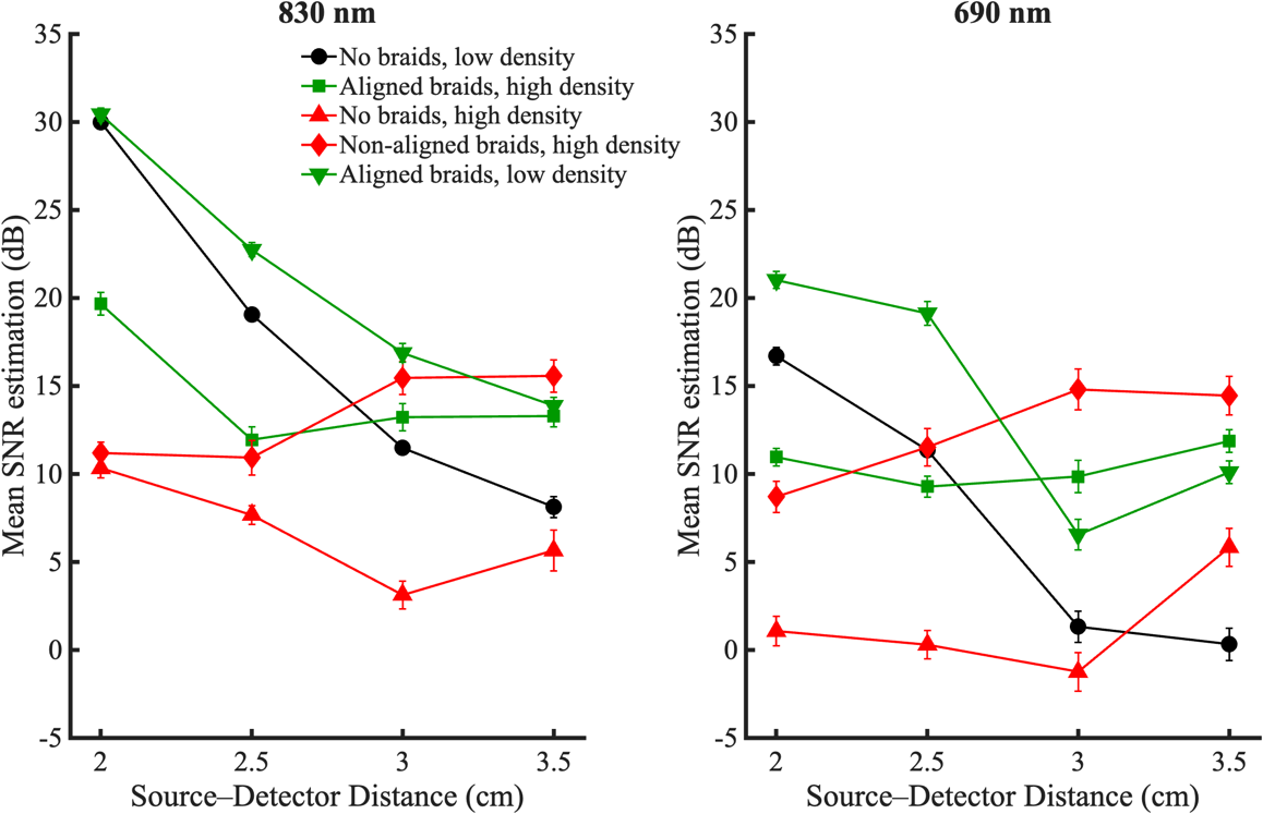
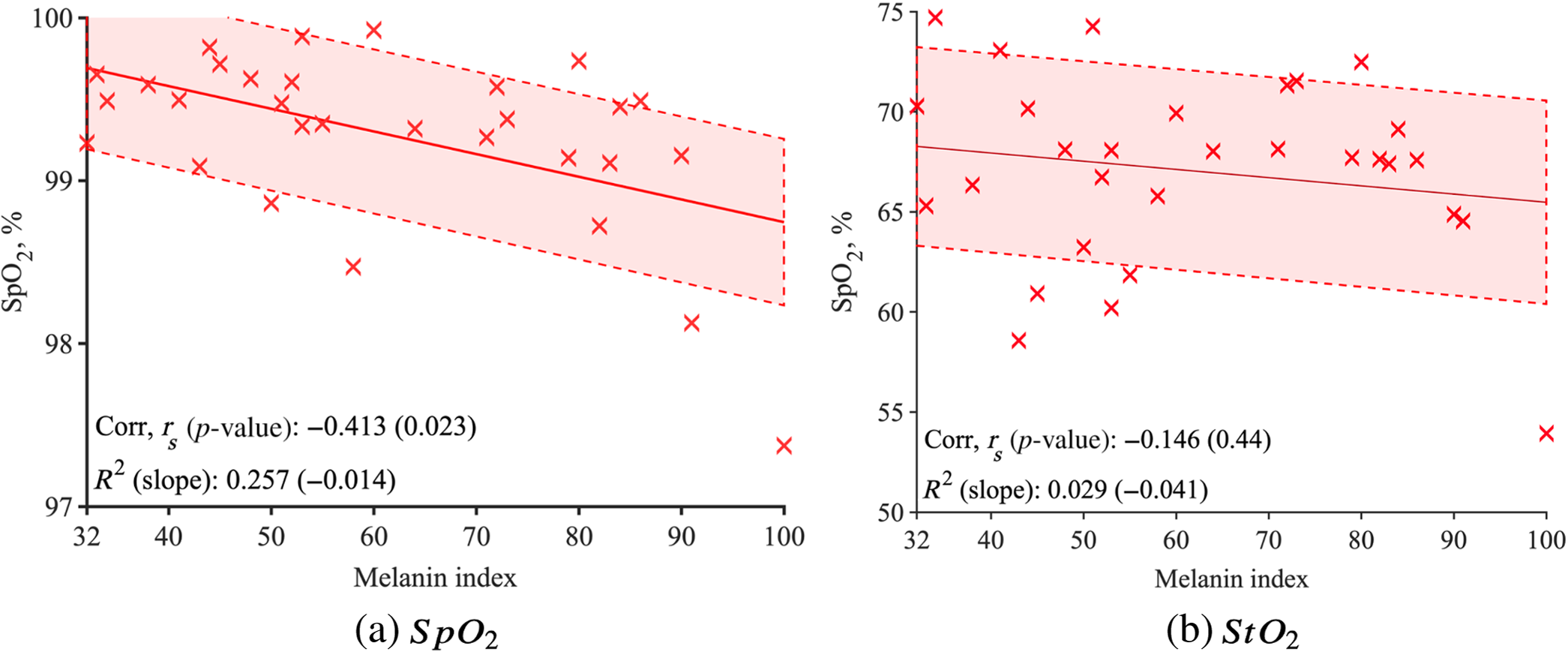
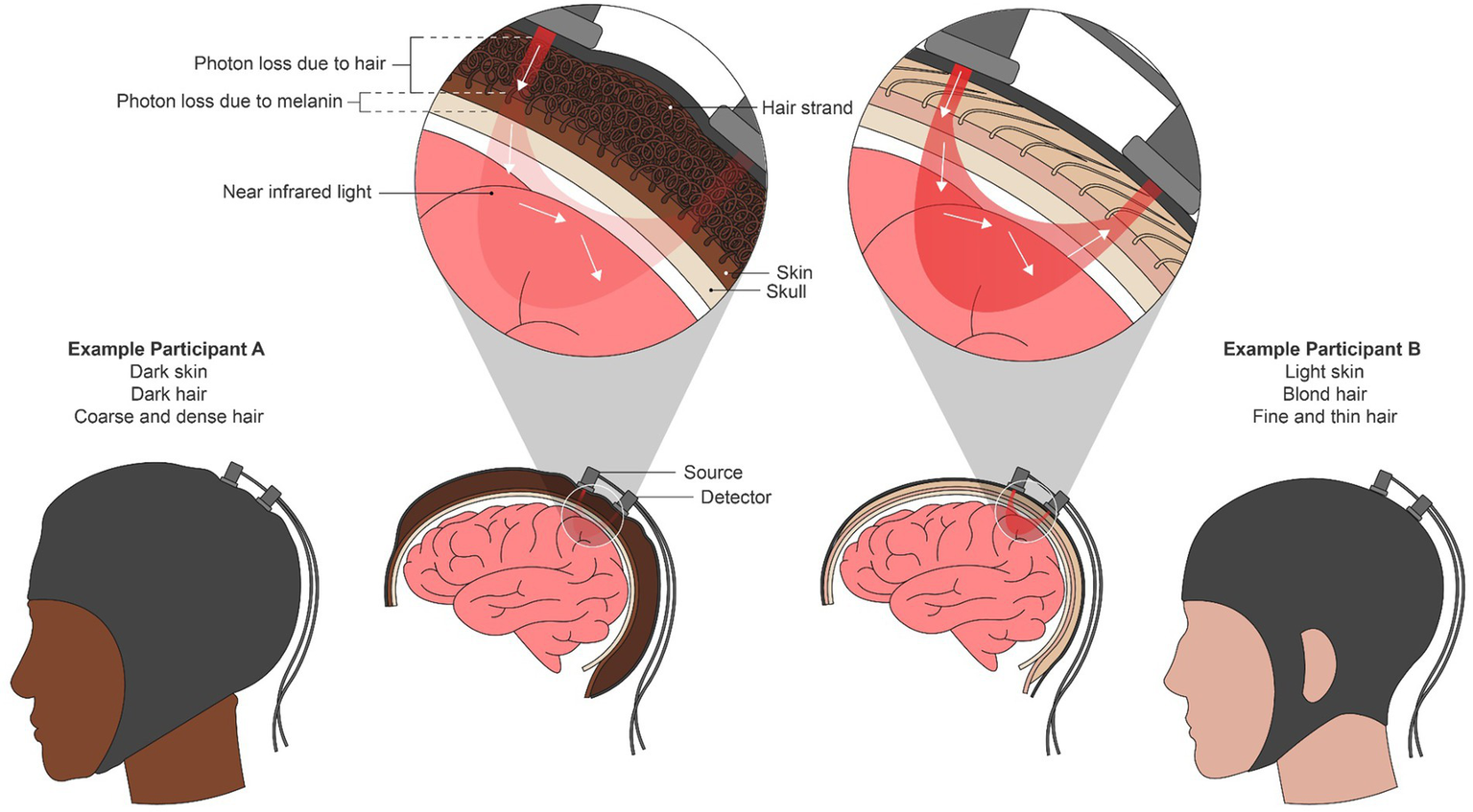
We design and evaluate neurotechnology and optical sensing tools that work across skin tone, hair type, and clinical context.Our team is comprised of talented students, postdoctoral fellows, research associates and faculty across three labs — Biophotonics Lab, Grover Lab and Wood Neuro Research Group. This research project focuses on identifying and eliminating systemic racial, physiological, and physical biases inherent in wearable medical and neuroimaging devices
Collaborators
Jana Kainerstorfer, Ph.D., Pulkit Grover, Ph.D., Jasmine Kwasa, Ph.D., CMU Pittsburgh
Assane Gueye, Ph.D., Carine Pierrette Mukamakuza, Ph.D., CMU Africa
Seif Eldawlatly, Ph.D., American University of Cairo
Dr. Charles Mudenge, University of Rwanda
Elsie Kaufmann, Ph.D., University of Ghana
Founding Papers
Duong, A., Roy, S., Meinert-Spyker, E., Cao, J., Kwasa, J., Kainerstorfer, J., Grover, P., and Wood, S.
Novel Optode Sensor Development for Functional Near Infrared Spectroscopy Systems and its use in Dark, Coarse, and Curly Hair
Proc. SPIE 13834, Clinical and Translational Neurophotonics 2026, 138340H (5 Mar 2026)
Roy, S., Wu, J., Cao, J., Disu, J., Bharadwaj, S., Meinert-Spyker, E., Grover, P., Kainerstorfer, J. and Wood, S.
Exploring the Impact and Influence of Melanin on Near-Infrared Spectroscopy Measurement
Journal of Biomedical Optics 29(S3), S33310 (25 September 2024).
Kwasa, J., Peterson, H., Jones, L., Karrobi, K., Parker, T., Nickerson, N., and Wood, S.
Demographic Reporting and Phenotypic Exclusion in fNIRS.
Frontiers in Neuroscience, Brain Imaging Methods. 2023, May.
https://www.frontiersin.org/articles/10.3389/fnins.2023.1086208/full
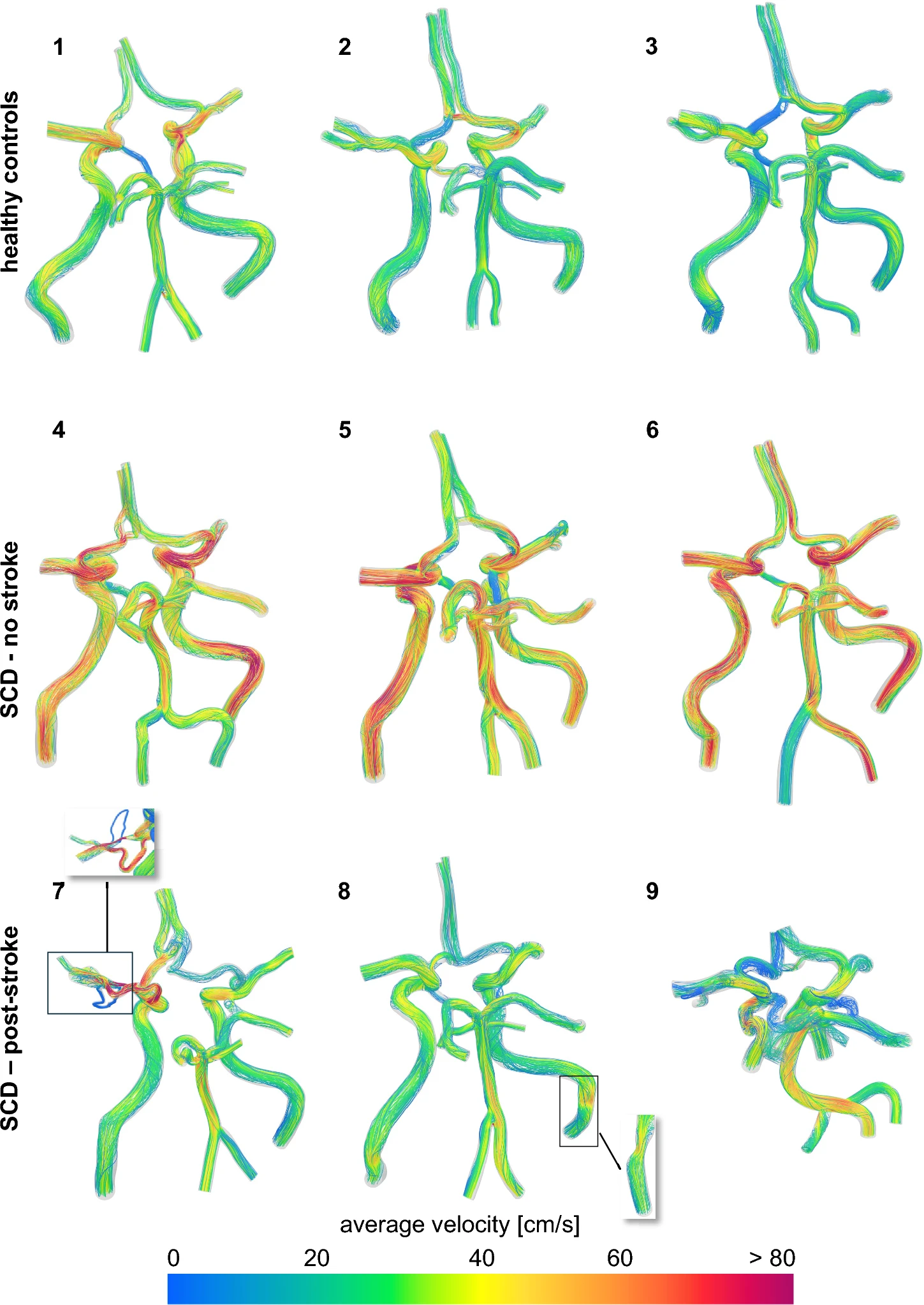
Patient-specific cerebral hemodynamics in sickle cell disease
We use patient-specific vascular imaging and computational fluid dynamics to understand how sickle cell disease changes blood flow in the brain and how those changes may relate to stroke risk.
In a close collaboration with Biomedical Flows Simulation & Multiscale Modeling lab, this project utilizes high-resolution MRI datasets to construct patient-specific computational fluid dynamics (CFD) models
Collaborators
Noelia Grande Gutiérrez, Ph.D., CMU Pittsburgh
Enrico M. Novelli, M.D., UPMC
Tamer S. Ibrahim, Ph.D., University of Pittsburgh
Brain networks, cognitive and behavioral neuroscience
Our research goal is to study the cortical and subcortical regions of the brain responsible for various cognitive functions such as decision-making, attention, and pain perceptions. We are especially interested in the role demographics such as race, gender, and cultural experience play in neural representations and discover any neural variations. Furthermore, we also want to examine the underlying process and identify any variations when humans are compromised with neurological or neurovascular diseases. Historically, the literature examining these questions has been scarce due to technological limitations, in which tools such as EEG and NIRS are noisier in specific populations than others. Thus, in addition to using state-of-the-art neural technologies to record brain activities, we have developed novel hardware and software to combine with the existing technologies to help answer these questions. We created simple and complex tasks designed to understand how neural circuits compute information and map out the organizational structure of the brain; and we further interpret the results due to demographics and disease.
Collaborators
Pain phenotyping and objective SCD pain biomarkers
We study how people with sickle cell disease describe pain and how those pain phenotypes relate to brain network activity.
Our group investigates the neurological markers of chronic pain in adults with sickle cell disease by mapping brain connectivity through the following methods:
-
Cortical Signatures: Identifying how the brain processes the transition from standard sensation to pain.
-
Network Connectivity: Studying how nociceptive (physical injury) and neuropathic (nerve damage) pain descriptors correlate with activity in specific brain networks (like the default mode, salience, and somatosensory networks).
-
Predictive ML Tools: Leveraging machine learning and mobile tools (like Painimation) to identify predictive features of chronic pain phenotypes.
Collaborators
Benedict Alter, M.D., Keith Voigt, M.D., UPMC
Charles Jonassaint, M.D., Emory University
Papers
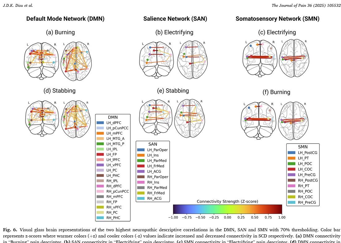
Disu, J. D. K., Jonassaint, C. R., Santini, T., Ibrahim, T. S., Novelli, E. M., & Wood, S., "Nociceptive and Neuropathic Pain Descriptors in Adults with Sickle Cell Disease are Associated with Overlap Activity in the Default, Salience and Somatosensory Networks. The Journal of Pain, 2025, 105532, August, Volume 36, https://doi.org/10.1016/j.jpain.2025.105532.
Imaging Disease Modifying Therapies in SCD
This multi-site study combines imaging and physiological monitoring to evaluate how disease-modifying therapies improve cerebral blood flow, oxygenation, and cognitive function in adults with sickle cell disease.
This is a multi-site study across Pittsburgh, PA and Nigeria—in close collaboration with Dr. Julia Xu's research team at UPMC—to evaluate the real-world effects of disease-modifying therapies on adults with sickle cell disease. Having successfully recruited roughly 100 participants across the study sites, this project represents one of the few international, multimodal imaging and rheological datasets of its kind
Studies: ACHiEvE-SCD, VASC
Relevant Proceedings:
- Momoh, S., Tukakira, J. Meinert-Spyker, E., Candra, L., Ibrahim, TS, Novelli, E.M., Wu, M., Wood, S., Xu, J., “Sex-related differences in silent cerebral infarction burden among adults with sickle cell disease,” Blood, Volume 146, Supplement 1, 2025 https://doi.org/10.1182/blood-2025-1193.
- Jallepalli, D., Roy, S., Meinert-Spyker, E., Xu, J., Wood, S. “Cerebral and Peripheral Microvascular Function in Sickle Cell Disease: Blood Flow and Oxygen Extraction via Diffuse Correlation and Near-Infrared Spectroscopy,” Blood, Volume 146, Supplement 1, 2025, https://doi.org/10.1182/blood-2025-2937.
- Kiriza, N., Abdelmohsen, L., Meinert-Spyker, E., Tukakira, J., Oluwole, F., Adiat, A., Disu, J., Mossazghi, N., Jonassaint, J., Olesugun Joseph, D. A., Osunkalu, V., Oluwole, O., DeCastro, L., Novelli, E., Adeyemo, T., Wood, S., Xu, J., “Raising hemoglobin level with erythropoietin improves cerebral but not muscle tissue oxygenation in patients with sickle cell disease,” Blood, Volume 146, Supplement 1, 2025, https://doi.org/10.1182/blood-2025-2964.
- Abdelmohsen, L., Mossazghi, N., Roy, S., Disu, J., Meinert-Spyker, E., Saber, C., Xu, J. Z., Wood, S., “Assessing the Impact of Cognitive Load on Resting State Tissue Oxygen Saturation in Adult Patients with Sickle Cell Disease,” Blood, Volume 144, Supplement 1, 2024, https://doi.org/10.1182/blood-2024-208977.
- Disu, J., Abdelmohsen, L., Mossazghi, N., Meinert-Spyker, E., Saber, C., Xu, J. Z., Wood, S., “Dynamic Cerebral Autoregulation and Blood Pressure Reveal the Severity of Autoregulatory Dysfunction in Sickle Cell Disease,” Blood, Volume 144, Supplement 1, 2024, https://doi.org/10.1182/blood-2024-210312.
- Saber, C., Meinert-Spyker, E., Abdelmohsen, L., Philips, B., Disu, J., Mossazghi, N., Kiriza, N., Nouraie, S., Oluwole, O., DeCastro, L., Straub, A., Novelli, E., Wood, S., Xu, J., Z., “Higher Hemoglobin Level Correlates with Increased Tissue Oxygenation in a Comprehensive Profile of Hemorheology in Sickle Cell Disease,” Blood, Volume 144, Supplement 1, 2024, https://doi.org/10.1182/blood-2024-203346.
Collaborators
Julia Xu, M.D., Busola Oluwole, M.D., UPMC
Titi Adeyemo, M.D., Lagos University Hospital
Research Partners
Clinical Collaborators
- UPMC Adult Sickle Cell Disease Program
- UPMC Anesthesiology
University Partners
- University of Pittsburgh
- Lagos Teaching Hospital
- University of Rwanda
- Emory University
- University of Ghana
- American University of Cairo
Imaging Collaborators