Study Reveals First Fine Structure of Complete Vertebrate Brain
Findings could lead to better understanding of human brains

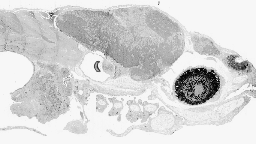
Thousands of electron microscopic images viewed on edge are combined to recreate the eye, ear, brain, nose and several vertebrae of a zebrafish.
Scientists have generated the first 3D map of a vertebrate brain that shows all the connections between nerve cells. The work, on zebrafish larva, holds promise in explaining how these comparatively simple brains work, and may offer insight into how more complex brains such as those of humans work as well. An international collaboration including the Pittsburgh Supercomputing Center (PSC) reported their initial results on May 10 in the journal Nature.
PSC is a joint program of Carnegie Mellon University and the University of Pittsburgh.
Zebrafish larvae are small and transparent and have been studied by researchers to understand how their tiny brains generate behaviors, according to principal investigator David Hildebrand. Working in the laboratories of Florian Engert and Jeff Lichtman at Harvard University, Hildebrand sliced the front quarter of the zebrafish larva into more than 18,000 slices and then created electron microscopic images of these slices.
The physical process of slicing inevitably distorted some of the slices. Co-author Art Wetzel at PSC helped to recombine the distorted images to reconstruct the brain in three dimensions. Wetzel used SWiFT (Signal Whitening image Fourier Transform) software he developed as part of PSC's involvement in the National Center for Multiscale Modeling of Biological Systems. SWiFT gave the scientists the ability to handle distortions and defects stemming from tissue variations, compression of slices and image distortions caused by the electron microscope's inner workings.
Thanks to Wetzel's work, the scientists collected more than 4,900 gigabytes of data in the process and fully or partially traced the path of about 2,500 nerve cells and their axons - the long tails the cells use to connect with other nerve cells. The investigators followed 805 of these nerve cells over the entire length of their axons through the brain.
"Our goal was to develop techniques that allow researchers to examine the morphology and circuit connectivity of any neuron in the brain of a larval zebrafish at about five days after fertilization," Hildebrand said. "This is when interesting zebrafish behaviors such as hunting emerge, giving us the opportunity to ask how circuits of neurons parse incoming information from the environment to generate useful behavioral outputs."
One early finding is that certain nerve fibers on one side (left or right) of the fish brain have twin fibers on the other side. The organization of axons within these nerves on each side followed nearly mirror-image paths. While the scientists do not know exactly what this means, they suspect it may have something to do with a pre-programmed brain development process. This also could be an important clue for a number of inborn behaviors fish follow.
It is not clear whether nerve cells in the human brain, which develop slowly and change greatly throughout life, will have the same degree of left/right symmetry.
"What makes the zebrafish such a spectacular system is that the alternatives in other organisms for deriving wiring diagrams are limited to a tiny, tiny part of a much larger brain, and so don't offer the opportunity to study the full range of an organism's behavior," Engert said. "Nobody previously had dared to think of doing this kind of work in a whole brain."
"Nobody previously had dared to think of doing this kind of work in a whole brain." - Florian Engert