Nano-CT: Xradia UltraXRM-L200
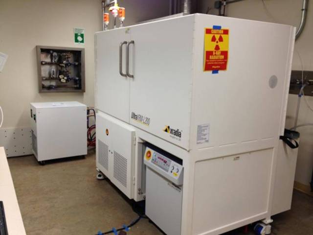
The XCTF features a Xradia UltraXRM-L200 nano-CT as shown below. The UltraXRM-L200 has the following features:
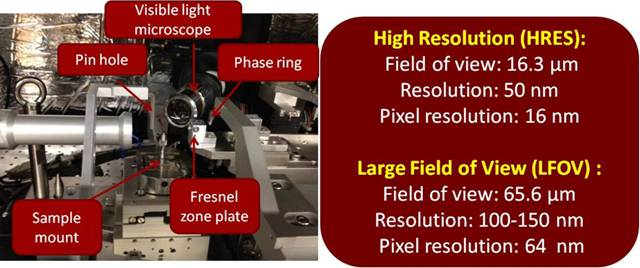
- Two levels of resolution- (a) 50 nm resolution (16 nm pixels) for a 16 µm field of view & (b) 150 nm resolution (65 nm pixels) for a 65 µm field of view;
- Conventional absorption contrast imaging mode at 8 keV. In this mode, the contrast is generated by the X-ray absorptivity of the sample (a function the local atomic number, Z, and density)
- To enhance imaging of low-contrast, soft materials (e.g., carbon and organic materials) with low Z, the nano-CT has a Zernike phase contrast mode. In this mode, a gold phase ring is used to image the phase shift of the X-ray beam going through the sample. This mode enhances the imaging of material boundaries in materials that are typically difficult to image, such as polymers and soft tissue.
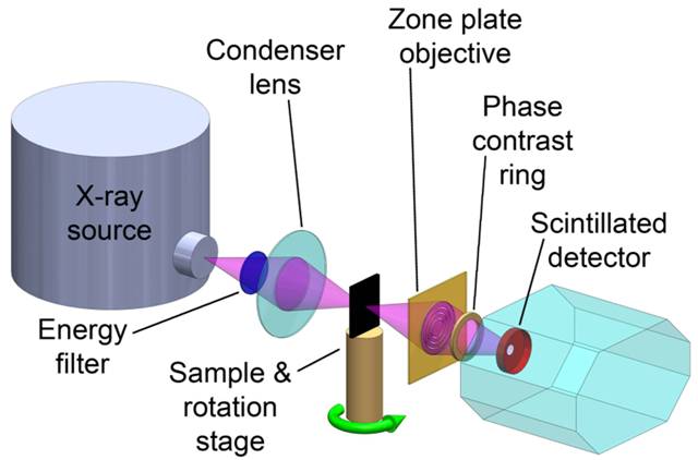
The UltraXRM-L200 achieves its resolution using a laboratory X-ray source (rotating copper anode) that emits X-ray with a photon energy level of 8 keV. As the schematic below shows, the X-ray beam passes through a mono-capillary condenser lens that uses grazing incidence reflection to efficiently focus the X-rays on the sample. This efficient condenser is key to using a laboratory X-ray source rather than a synchrotron beam. After passing the sample, the X-rays are focused onto the detector using a Fresnel zone plate objective. The zone plate objective consists of high aspect ratio concentric gold rings. The maximum resolution of an X-ray transmission microscope is related to the minimum spacing of the gold rings. The 35 nm spacing of the high resolution zone plate in the UltraXRM-L200 yields a theoretical Rayleigh criterion resolution of 43 nm. After the zone plate, the X-ray beam passes by a gold phase ring for Zernike phase contrast (if in phase contrast mode). The ring phase shifts X-rays not diffracted by the sample, causing interference between the undiffracted X-rays and those diffracted by the sample and resulting in a negative phase contrast image. Subsequently, the X-rays intercept a scintillation screen coupled to a CCD detector.
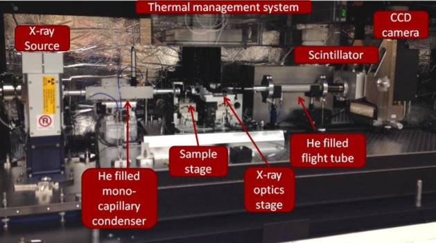
Below is an image of the layout within the UltraXRM-L200.
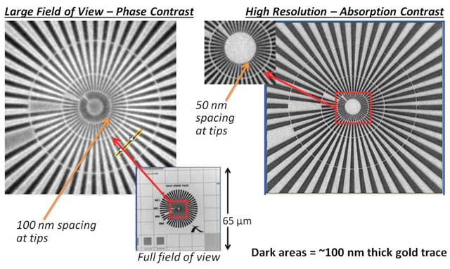
Below are radiographs obtained using the UltraXRM-L200 in the XCTF. The images on the left are from imaging in the large field of view (65 um) mode with Zernike phase contrast turned. Nearly 100 nm resolution was obtained. The images on the right are for the high resolution mode with absorption contrast and demonstrate the 50 nm resolution.
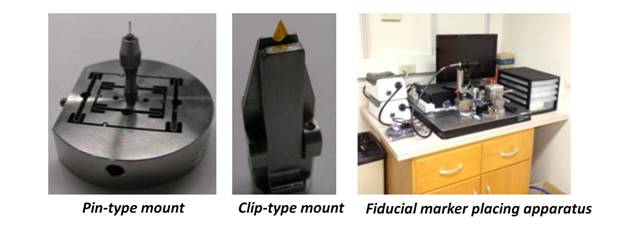
Samples can be prepared and mounted in a variety of methods. The sample holder area is exposed to the ambient air, but samples can be placed in sealed chambers (e.g., Kapton tubes). The image below shows the two sample mount types for the nano-CT. The samples can be mounted by epoxy or by other means (FIB Pt weld) to the tip of a pin and placed in the pin-type sample mount, or films and similar objects can be placed in the clip-type sample mount. In the 50 nm high resolution mode, it is reccomended that a 1-2 um gold particle fiducial marker is placed on the sample, which can be done with the apparatus in the facility.
For the best reconstruction quality, it is recommended that samples be small enough to fit within the field of view at all projection angles (<16 um wide for 50 nm resolution, <65 um wide for 150 nm resolution). However, in some cases, high quality can be obtained with larger sample - depends on sample material and geometry. In addition, high Z materials cause high attentuation and may require the sample to be even smaller in order to get the desired contrast. Brian Patterson at Los Alamos National Laboratory has generated and posted this useful periodic table containing 1/e penetration depths for the Ultra-XRM's 8 keV photon energy.