Tiny Dots on a Big Mission
Surgeons are continually seeking better imaging techniques for cancer detection. One particular area that's been challenging for them is capturing brain cancer on film.
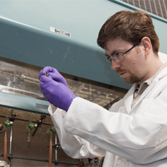
"Photographing brain cancer isn't easy," explains Carnegie Mellon's Marcel Bruchez, developer of quantum dots, or Q-dots for short. "Q-dots emit infrared light, outlining the tumor and allowing it to be photographed."
How it works: White blood cells act like taxis, carrying the fluorescent Q-dots to wherever there is infection or inflammation.
With a large enough dose, the Q-dots outline the tumor and neurosurgeons are able to see its size, shape and location — information that could help them remove tumors with less damage to the surrounding healthy brain tissue.
Dr. Steven A. Toms, a neurosurgeon in the Cleveland Clinic's Neural Science Institute, is helping Bruchez refine the procedure, which could later be applied to tumors elsewhere in the body.
The optical equipment necessary would cost between $10,000 and $50,000 — significantly less than the millions of dollars required for the MRI imaging being used for brain cancer.
Bruchez holds 16 patents and has received prestigious awards for his work, including MIT's TR100 in 2004, which recognizes 100 innovators under age 35 who are transforming technology.
Related Links: Read More | Dept of Chemistry | MCS
Homepage Story Archives