Shining a Light on Tissue Health
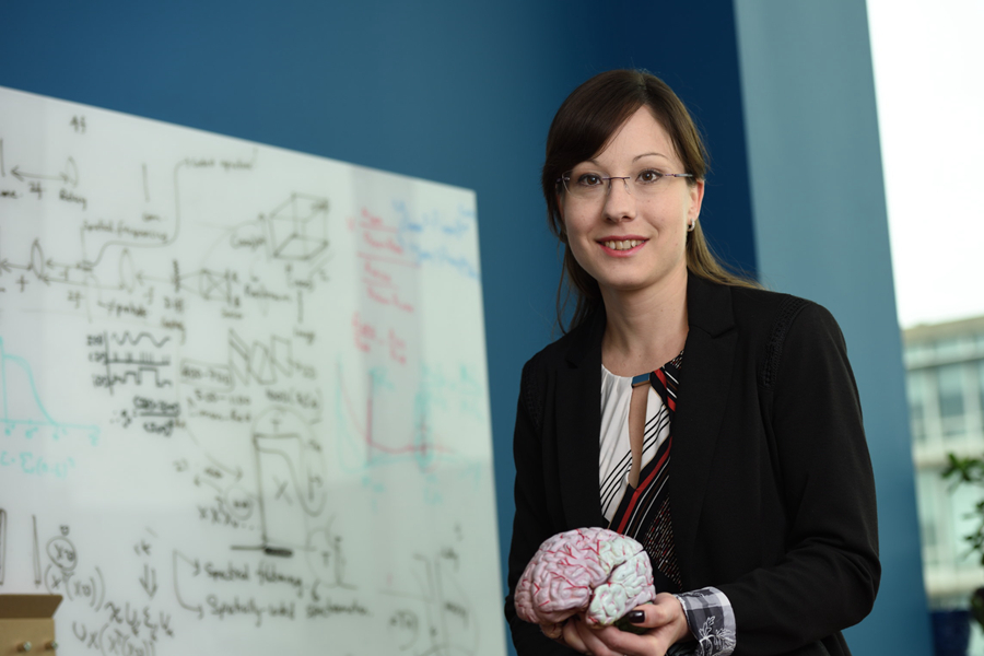
Professor Jana Kainerstorfer is using non-invasive optical methods to monitor and detect human tissue disorders
Early detection of disease is one of the keys to gaining a positive outcome for patients. However, current methods of monitoring changes inside the human body are at best, expensive — and, at worst, highly invasive. In her Biophotonics Lab in BME, Professor Jana Kainerstorfer is developing new, light-based methods for assessing the health of living tissues inside the human body.
The human brain represents an incredibly complex organ and computational system. But, when something goes wrong in the brain, whether caused by an injury or disorder, the effects are largely invisible. One of the goals of Prof. Kainerstorfer’s research is to monitor cerebral disease more effectively and routinely at the bedside.
“By using near-infrared lasers, we can gain a clear view of multiple centimeters of tissue inside the human body in a non-invasive, painless way,” explains Kainerstorfer. “We hope to detect disease at an early stage, or notice changes in tissue over time via continuous monitoring.”
Applications of Prof. Kainerstorfer’s novel methods include measuring pressure changes inside brain tissue following a stroke or traumatic head injury. “By implementing this technology at a patient’s bedside, we hope to be able to reduce the use of CTs, MRIs and other costly imaging procedures — while also providing long-term, continuous imaging, versus a one-time perspective,” says Kainerstorfer.
Such research is not possible without collaborations, such as with Matthew Smith, a professor of ophthalmology at the University of Pittsburgh, with whom she developed the non-invasive intracranial pressure sensor using the near-infrared imaging technology. Clinical translation of such methods is possible due to partnering with Dr. Tyler Kabara, a neurosurgeon at Children’s Hospital of Pittsburgh.
In addition to her efforts in clinical translation of near-infrared imaging methods, Prof. Kainerstorfer is also part of a team of Carnegie Mellon researchers that aims to develop high-performance, bi-directional brain-machine interfaces. The team has recently been awarded a $19.48 million grant from the Defense Advanced Research Projects Agency (DARPA), with the team hoping to use noninvasive neural interfaces to harness novel concepts in physics, biology, and engineering through electricity, ultrasound, and light. The team of researchers is co-led by Professors Pulkit Grover, Maysam Chamanzar and Kainerstorfer. Other investigators in the project include BME faculty Gary Fedder, Aryn Gittis, Alison Barth, Shawn Kelly, and Bin He, together with collaborators at University of Pittsburgh, Battelle Memorial Institute, and the Air Force Research Lab.
“While there is no medical school at Carnegie Mellon, BME is part of a vibrant, integrated, collaborative health care research community here in Pittsburgh,” notes Kainerstorfer. “I’m fortunate that BME makes it so easy to identify external experts and partner with them. It’s really the perfect environment for my work, which is very multidisciplinary in nature — and relies on real-world testing in a clinical setting.”
Kainerstorfer, who joined BME in 2015, also enjoys the opportunity to work with student researchers in BME. “The students at Carnegie Mellon are really brilliant, and I love seeing them progress and grow over time,” she says. “It’s wonderful to have a collaborative team where we come up with this innovative idea that has the potential to change lives — then we get to see it proven. It’s very rewarding, and I’m grateful for the freedom and support I enjoy here.”