A new platform for neural recording from large brains
In our quest to understand the human brain, there are many obstacles standing in our way. Not only are the anatomy and structure of the brain incredibly complex and difficult to study, but there is also a fine line between taking safe, accurate readings and causing irreparable damage.
A team led by Maysam Chamanzar, an assistant professor of electrical and computer engineering and biomedical engineering, has been awarded an R01 grant from the National Institutes of Health (NIH) to develop a novel neural interface made from stainless steel for high-density neural recording, making the brain readings much safer than before.
These stainless steel neural probes, which Chamanzar calls “steeltrodes,” will minimize the risk of breakage during surgery—and thus the risk of residue being left behind in the brain—a potential problem with the current silicon neural probe technology.
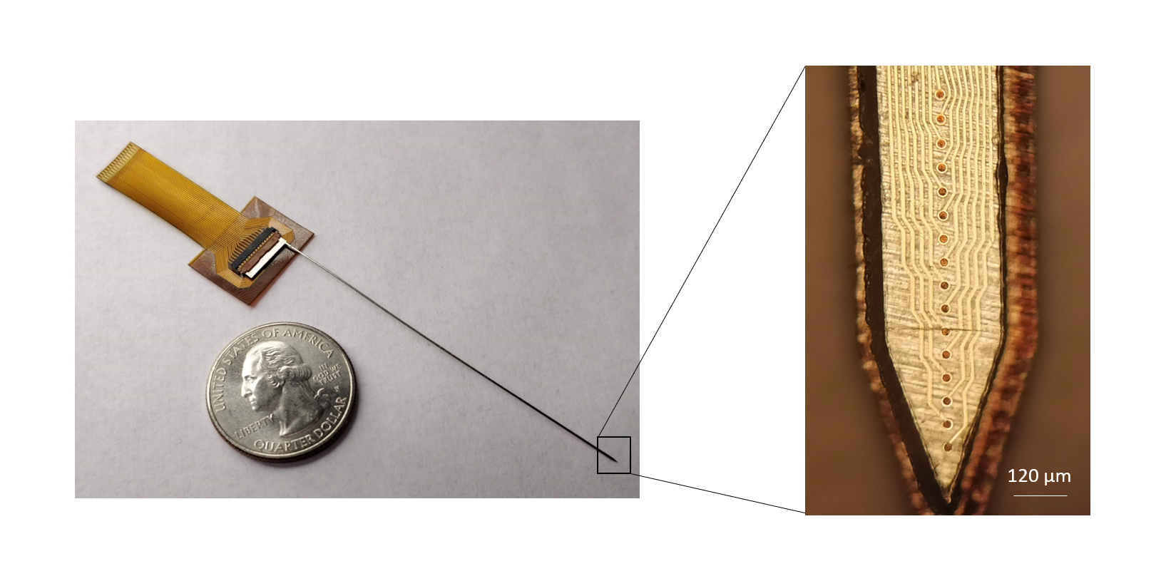
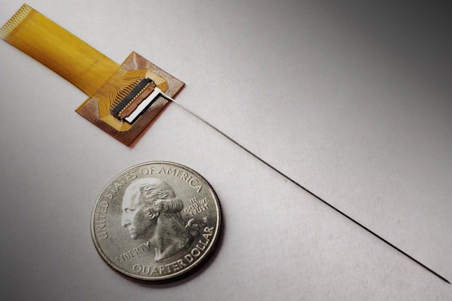
Source: College of Engineering. A steeltrode compared to a quarter for scale.
Implantable neural probes are in high demand for clinical applications, such as recording brain activity during brain surgery. Such neural interfaces are also needed for chronic, longer-term monitoring of brain function, for example, to help neurosurgeons localize the sources of epileptic seizures in the brain. Some of these neural probes are permanently implanted in the brain for deep brain stimulation (DBS), in order to mitigate conditions such as Parkinson’s disease.
In the future, Chamanzar’s novel neural interfaces will allow these therapeutic interventions to be performed safely and more accurately. Moreover, the novel technology may enable the development of new groundbreaking treatments and therapeutic interventions for neurodegenerative diseases.
Until now, implantable clinical neural interfaces have been prohibitively large, and only provide a very limited number of channels (electrodes) for neural recording and stimulation. What’s more, the specialized methods used to manufacture these neural probes make them costly and largely inaccessible to a broad patient base.
High-density needle-like neural probes—with a large number of channels in relation to their very small form factor—have already been designed for studying neural circuits in small animal models such as rodents. By leveraging microfabrication techniques developed to build electronic chips, researchers have recently been able to develop high-density neural probes mainly using silicon as the material of choice.
But silicon is a very brittle material, and when made into a shape so thin and needle-like, the chance of it breaking under pressure is very high. Even a small force on these probes can cause them to shatter, making them very difficult to work with. To address this problem, Chamanzar and his team are looking to a different material, one already widely used in surgical applications.
“After a series of discussions with neurosurgeons and understanding real needs in the application domain, we came up with the idea of using stainless steel to make ultra-high-density neural probes, because stainless steel is very resilient, and it cannot break easily,” explains Chamanzar. “At worst, stainless steel neural probes will bend, rather than breaking and leaving behind residue.”
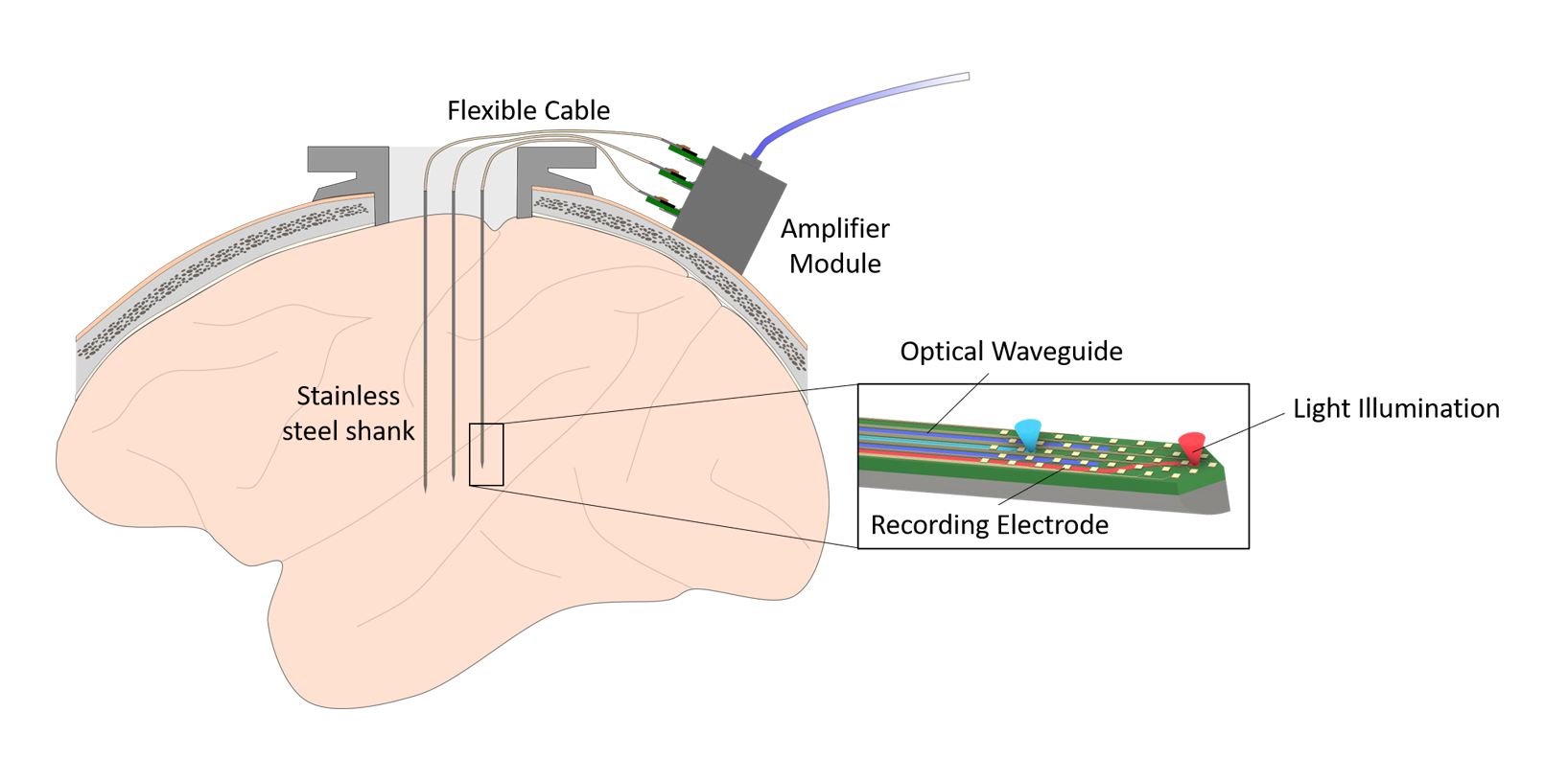
Source: College of Engineering. A diagram of steeltrodes interfacing with the brain.
Brain circuits are very dense and complex. Therefore, a large number of recording channels are desired to get a more realistic picture of how they function, or sometimes dysfunction. But adding more channels presents its own set of challenges.
“Our goal is to make a bidirectional brain interface, with a path towards clinical applications in humans,” says Chamanzar. “We would like to make the implantable device very thin and long to minimize damage to the brain tissue, while reaching deep regions. The long aspect ratio makes the design challenging, especially when you want to add a large number of recording sites.”
In addition to electrical recording and stimulation, Chamanzar is planning to integrate optical channels on his neural probes to provide visual access to the brain to enable imaging, as well as optical manipulation of neural circuits.
In this project, the optoelectrical steeltrodes will be developed for testing on large non-human primates before human translation.
“The opto-steeltrodes open up a number of unique experimental opportunities for neural recordings and modulation in large primates, and ultimately also in humans,” says Tobias Teichert, an assistant professor of psychiatry and bioengineering at the University of Pittsburgh and a collaborator on the project. “I am particularly excited about the opportunity to combine layer-specific neural recordings with layer-specific optical interventions.”
Chamanzar’s team will work closely with Teichert’s research group, as well as those of Pitt faculty Susanne Ahmari and William Stauffer, to test and demonstrate the steeltrodes for the study of auditory systems in large animals using electrophysiology recording and optogenetic stimulation. Currently, very few technologies exist to allow researchers to study the neural systems of large animals, making this novel neural interface a huge leap forward in an underdeveloped field of research.
Chamanzar’s interdisciplinary research is at the interface of engineering and neuroscience. His lab is committed to bridging the gap between neuroscience and engineering through designing advanced neural interfaces that can address outstanding needs in neuroscience studies and clinical practices.