
'Reporter' Technology
Carnegie Mellon University biologists have developed an MRI-based technique that allows researchers to non-invasively follow neural stem cells in vivo.
The recently patented technology could be used to further the study of neural stem cells and inform the development of new treatments for brain injury caused by trauma, stroke, Parkinson's disease and other neurological disorders.
The findings, authored by CMU Associate Professor of Biological Sciences Eric Ahrens and Biological Sciences postdoctoral student Bistra Iordanova, are published online in the journal NeuroImage.
Legend had it that once a brain cell dies, it's lost forever. Neuroscientists now know that this is purely myth, having proved that the brain is constantly producing new neurons.
These neural stem cells are born deep in an area of the brain called the subventricular zone. As time goes on, the cells, also called neuroblasts, make their way to other areas of the brain where they mature into functioning neurons.
The brain's ability to regenerate its cells is of great interest to scientists.
"If we could better understand the molecular migratory signals that guide neuroblasts, we could try to redirect these cells to areas of the brain harmed by stroke or traumatic brain injury.
With this information, scientists might be able to one day repair the brain," said Ahrens, who also is a member of the Pittsburgh NMR Center for Biomedical Research.
Studying cells in a living brain is problematic. Common forms of in vivo cell imaging like fluorescence and bioluminescence rely on light to produce images, making them unsuitable for viewing neuroblasts buried deep beneath the skull and layers of opaque tissue.
Until now, scientists had only been able to study neuronal stem cells by looking at slices of the brain under a microscope.
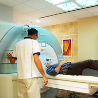
Ahrens was able to surmount this problem using MRI technology.
Rather than light, MRI uses magnets to create high-resolution images. A typical MRI scan uses a magnetic field and radio frequency pulses to cause the hydrogen protons found in the body's water molecules to give off signals. Those signals are converted into a
high-resolution image.
At the foundation of this work is a technology Ahrens developed. (Read more in a 2005 issue of Nature Medicine.) Recently, Carnegie Mellon received a patent for this 'reporter' technology.
Ahrens hopes to continue to develop the technology in order to allow researchers to better understand neuronal stem cells and how neurons regenerate.
Ahrens also plans to use the reporters to improve clinical trials of cell-based therapies. By incorporating the reporter into the cells before implantation, researchers would be able to find the answer to a number of critical questions.
"Where do these cells go, days, weeks and months later? How do we know that they've grafted to the right cells? Or have they grafted in the wrong place? Or died?" Ahrens asked. "The reporter can show us the answers."
The National Science Foundation and National Institutes of Health funded this research.
CMU has been a leader in the areas of brain science, psychology and learning research for many decades.
With the launch of the university's new brain, mind & learning initiative, CMU plans to become an even bigger player in these fields, while distinguishing ourselves from other brain research programs.
Related Links: Eric Ahrens | Dept of Biological Sciences | Brain, Mind & Learning